The authors review three different bodies of evidence having to do with face processing: behavioral studies (i.e., comparing autistic and neurotypical people's accuracy in identifying/distinguishing individual faces, or classifying facial expressions, from videos or photos; faces might be those of strangers, or of people known to the subjects, and might also be turned upside down or partially covered up in some experiments), electrical and magnetic evoked potential studies (i.e., doing EEGs or MEGs of people as they look at images of faces), and brain-imaging studies (doing fMRIs or PET scans of people as they look at images of faces).
The behavioral literature on how autistic people perceive faces shows that, though we are not necessarily any worse at identifying faces, or facial expressions*, we tend to go about it using different methods.
We look at different parts of the face: some studies --- like Joseph and Tanaka, 2003 (full text here); and Klin et al., 2002 --- find that we spend a lot more time looking at the lower parts of faces, particularly the mouth, while others --- among them van der Geest et al., 2002 (full text here); Dalton et al., 2005 (full text here); Lahaie et al., 2006 (full text here); Bar-Haim et al., 2006 (full text here) and de Wit et al., 2008 (full text here) --- find that we don't direct our gaze** to the lower face any more often than nonautistic people do. Some other studies have found that we attend to lots of different visual details --- both within the face and outside it --- that neurotypical subjects ignore.
Examples of studies finding a greater degree of attention to "irrelevant" visual details: Pelphrey et al. (full text here) found that the five*** autistic men they studied looked more often at "nonfeature areas of the face", like the hairline, chin, jawline, ear, temples, etc., while the five neurotypical men who served as a control group mostly looked at the triangle-shaped region in the center of the face, encompassing the eyes, nose and mouth. More recently, de Wit et al. also found this to be the case: their autistic subjects spent less time than controls did looking at either the eyes or the mouth --- about half of all their viewing was directed at areas of the face other than eyes, nose or mouth. Also, Dalton et al. found that, while the autistic young men they studied did not focus particularly on the mouth, or on any other particular feature, they definitely avoided the eyes.
Since Dalton et al. were monitoring brain activity along with gaze direction, they noticed intense activity in the amygdalae and orbitofrontal cortices of the autistic subjects whenever they focused on the eyes of the faces they were supposed to be looking at. This effect was totally absent in the nonautistic subjects, which led the authors to consider the possibility that maybe autistic people shy away from meeting people's eyes to spare themselves this nerve-wracking emotional stress:
Notably, the autistic group showed greater activation in the left amygdala and orbitofrontal gyrus than did the control group in response to the standardized emotional facial photographs, and greater right amygdala activation in response to the familiar and unfamiliar facial photographs. These are the areas associated with emotional processes, and these results suggest that processing of faces is associated with a heightened activation in affective central circuitry in autism. Together, these findings suggest that the increased activation in subjects with autism in the orbitofrontal cortex occurs specifically in response to the emotional content of the faces and not to faces in general, whereas activation in the amygdala is not specific to the emotional content of the faces but [is] a response to faces in general.This hypothesis --- that autistic people are actually hypersensitive, rather than oblivious, to the emotional content of faces, especially in the eyes --- gets some additional support from de Wit et al.'s main finding. They showed a group of autistic children and a group of typically developing children four photographs of faces --- two with "positive" emotional expressions (a calm face and a smiling face) and two with "negative" ones (a frightened face and an angry face) --- while tracking where the children directed their gaze. They found that both groups looked more at the eyes of the "negative" faces, which seems to me an indication that the autistic children were able to detect, and attend to, the socially threatening messages encoded in the angry and fearful faces. (However, the hyperarousal hypothesis does have some contradictory evidence in the findings of Pierce et al., 2004 --- they found no significant differences in amygdalar activity between autistic and nonautistic people looking at faces).
These findings indicate that the commonly reported hypoactivation in the fusiform gyrus during face processing in autism may be a function of how individuals with autism scan the face rather than a group difference in which area of the brain is used to process faces. ...
...
On the basis of these findings, we suggest that within the autistic group, eye fixation is associated with negatively valenced overarousal mediated by activation in limbic regions such as the amygdala. We propose that diminished gaze fixation within the autistic group may facilitate reduction of overarousal to social stimuli. According to this model, face-processing deficits in autism arise from hyperactivation in the central circuitry of emotion that produces hypersensitivity to social stimuli, leading to characteristic diminished gaze fixation, which in turn results in atypical activation of the fusiform gyrus. Additional studies including those involving direct measures of arousal (both self-reported and physiological) are warranted to further test this model.
This 2006 article by Dirk Neumann, Michael Spezio, Joseph Piven and Ralph Adolphs, published in Social Cognitive and Affective Neuroscience (and not cited by Jemel, Mottron and Dawson), might help shed some light on the issue of whether or not, and in what contexts, autistic people rely on "looking you in the mouth" to decipher socially salient information: they found no differences whatsoever between their autistic and nonautistic subjects' gaze direction and accuracy at face-reading, except when they asked them to interpret images of faces that had been digitally obscured, making the task of identifying the emotion pictured very difficult.
We asked high-functioning subjects with autism to identify emotional facial expressions, a task people with autism often perform normally. In line with previous findings, we also observed normal accuracy and normal face gaze, provided that whole upright faces were used as the stimuli. When the difficulty of the task was increased and only sparse regions of a face were revealed using the bubbles technique, we still observed normal performance levels in the autism group. However, their gaze onto these sparse facial features became dramatically abnormal. People with autism looked more often and longer (autism, 27.3%; control, 8.7% of all saccades) at the mouth.In other words, we can interpret faces one of two ways, one of which we are as good at using as nonautistic people are (i.e., the normal way, looking primarily at the eyes) and the other of which works even better, and seems to be autism-specific (i.e., looking primarily at the mouth). The study participants seemed to use the typical, eye-centered face-processing strategy as their default method, switching to the other only when the task became much more difficult, and the image from which they were to extract meaning very poorly visible.
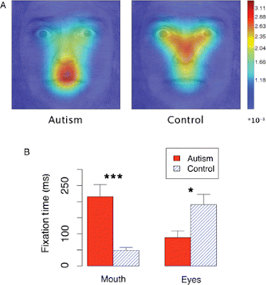 (Fig. 4 from Neumann et al., 2006. Top image is of areas of gaze fixation for the "bubbled" images; bottom image is a graph of how much time autistic vs. nonautistic subjects spent looking at eyes vs. mouth on "bubbled" faces)
Here's what Neumann et al. have to say about their findings:
Whatever the reasons may be for using one or another, it is certainly clear from these studies that autistic people have a wider range of face-perception strategies, the neuroanatomical correlates are unclear. So it seems to me that, unless a gaze-tracking apparatus is also used, and brain activity correlated to the part of the stimulus actually being viewed, as in Dalton et al. (2005), researchers hoping to pin down the neurophysiological underpinnings of autistic facial perception have no way of knowing that their experimental and control groups are actually doing the same task.Our results are consistent with earlier findings that autism subjects are impaired when judging complex social information from the eyes, but not from the mouth (Baron-Cohen, 1995), and that they rely more on information from the mouth for emotional judgments (Spezio et al., 2006). The differences in attentional processing suggest a possible general mechanism for the neurodevelopmental progression of impaired face gaze in autism (Dawson et al., 2005). We failed to observe a deficit in using low-level visual saliency cues, or bottom-up attention, while we found differences in top-down modulation for saccades made to the mouth. We propose that this evidence is consistent with a neurodevelopmental progression that begins in infancy with a failure in directing attention to the faces and more specifically to the eyes in a face, along with preservation of bottom-up attentional processing. This is consistent with the major deficits in social engagement rather than in nonsocial areas when autism is evident in early infancy (Kanner, 1943). Reported signs of lower social engagement in infants with autism include less interest in people (Volkmar et al., 2005) and less looking at faces (Osterling et al., 2002). The abnormal top-down attentional processing of faces may be due to abnormal reward circuitry (Dawson et al., 2005) or to abnormal circuitry for emotional salience (Schultz, 2005). Over the course of development, via learning, top-down attention may cause the propensity for mouth gaze in autism. This hypothesized mechanism adds to previous hypotheses about the causes of deficits in top-down attention to the face, and draws on our findings that bottom-up attention to the face is normal in high-functioning people with autism.
Another possibility is that the bubbles stimuli reveal an impairment in how attention is directed to local vs. global features. In contrast to earlier reports, we observed a normal eye gaze to whole faces. This could also be due to a social training program most of our autism subjects participated in. Despite their overall normal gaze to whole faces, the sparse bubbles stimuli might reveal an underlying impairment in deploying attention to local (vs. global) features (Dakin and Frith, 2005).
*Some of us are indeed "face-blind," or prosopagnosic. Prosopagnosia is not only seen in autistic people, though; it's a totally different phenomenon.
**You might be wondering how researchers can tell where their subjects are looking. There are several methods, but all the ones used in these studies seem to rely on some sort of head-mounted camera recording the infrared light that reflects off of a subject's corneas.
***The n for many of these studies is very small. I mentioned that for Pelphrey et al. (2002), n = 5; while that is the smallest sample size of any study I cite, most of the others aren't a whole lot bigger. Klin et al. (2002) have 15 autistic and 15 control subjects; Joseph and Tanaka have ~60 typically developing children and ~40 autistic ones; Neumann et al. have ten autistic subjects and ten NT ones; for van der Geest et al., the numbers are 16 autistic and 14 NT; Dalton et al. did two separate experiments, one involving eleven autistic subjects and twelve NT ones, the other involving 16 autistic subjects and 14 controls; Lahaie et al. have 16 autistic and 16 control subjects; and Bar-Haim et al. have twelve autistic and twelve typically developing subjects.
Dalton KM, Nacewicz BM, Johnstone T, Schaefer HS, Gernsbacher MA, Goldsmith HH, Alexander AL, & Davidson RJ (2005). Gaze fixation and the neural circuitry of face processing in autism. Nature neuroscience, 8 (4), 519-26 PMID: 15750588
Jemel, B., Mottron, L., & Dawson, M. (2006). Impaired Face Processing in Autism: Fact or Artifact? Journal of Autism and Developmental Disorders, 36 (1), 91-106 DOI: 10.1007/s10803-005-0050-5
Neumann D, Spezio ML, Piven J, & Adolphs R (2006). Looking you in the mouth: abnormal gaze in autism resulting from impaired top-down modulation of visual attention. Social cognitive and affective neuroscience, 1 (3), 194-202 PMID: 18985106
Pelphrey KA, Sasson NJ, Reznick JS, Paul G, Goldman BD, & Piven J (2002). Visual scanning of faces in autism. Journal of autism and developmental disorders, 32 (4), 249-61 PMID: 12199131
6 comments:
Jemel et al. (2006) was written in 2005 and published in January 2006. This explains why we did not cite Neumann et al. (2006), which was not accepted until September 2006.
In Lahaie et al. (2006), autistics, but not nonautistics, showed enhanced processing of information from eyes (in the form of significantly greater priming effects) compared to mouths.
Also it's worth mentioning that Pierce et al. (2004) found typical fusiform gyrus activity in autistics observing face images, to go along with the typical amygdala activity mentioned here. The proposals put forward by Dalton et al. (2005) would preclude this result (and some others).
When the results of experiments are so inconsistent with each other one has to wonder why?
One has to ask, what factors would one have to change in each case to bring consistency about.
What are the variables?
What if the variable was the concept of autism in each study itself, were the inconsistent thing in it all?
Well it is food for thought anyway.
@Michelle,
Yes, I noticed that Jemel, Mottron and your article was in the January issue of JADD, so I figured you probably wouldn't have seen Neumann et al. (2006) before sending your article in for review.
@Larry (you are Larry Arnold, aren't you?),
That's a good point; toward answering it, I can tell you what instruments each study used to diagnose autism in its participants:
- Joseph and Tanaka (2003): ADI-R
- Klin et al. (2002): ADI-R and ADOS; all participants also scored relatively high (average score = 101.3) on verbal IQ derived from Weschler scales WISC-III and WAIS-III and very low (average score = 46.7) on Vineland social-functioning scale VABS-E.
- van der Geest et al. (2002): ADI-R; all participants had fairly high Weschler IQs (average score = 92.8) and met criteria for DSM-IV Autistic Disorder or PDD-NOS
- Dalton et al. (2005): ADI-R; all participants also met DSM-IV criteria for Autistic or Asperger's Disorders, were verbally fluent, and had IQs (Stanford-Binet or Wide Range Intelligence Test) near 100.
- Pelphrey et al. (2002): ADI-R; all participants also met DSM-IV/ICD-10 criteria for autism and had fairly high Weschler (WAIS-R) IQs, with verbal IQ (average score = 117) somewhat higher than performance (average score = 86.5).
- Lahaie et al. (2006): ADI-R, ADOS-G, fairly high Weschler IQs (average score = 108.69, individual scores ranged from 93 to 130), with verbal and performance IQs not significantly different.
- Bar-Haim et al. (2006): DSM-IV Autistic Disorder, ADI-R, ADOS, relatively high WISC-III IQs (average score = 96.17)
- de Wit et al. (2008): ADI-R
- Pierce et al. (2004): ADI/ADI-R, ADOS; a mix of different combinations of Weschler IQ scores --- for most subjects, performance IQ was significantly higher than verbal (of the eight participants, only one had a higher verbal IQ), and full-scale IQ varied dramatically, from 55 to 104. One subject appears to be nonspeaking.
- Neumann et al. (2006): DSM-IV/ICD-10, ADI-R, ADOS
This is a start, anyway. To really answer your question I'll have to look into each of these scales in more depth, and see if use of any one of them over any other might be more likely to select for certain subtypes within autism.
(One thing I can tell you, right away, is that most of these researchers were dealing with verbal autistics; only one study, de Wit et al. (2008), mentioned having nonspeaking autistics in its sample, and that study did find differing gaze patterns between speaking and nonspeaking autistics.)
In Dalton et al. (2005), in the first experiment, one participant is described as "non-verbal" and two as having "minimal functional speech with pronounced echolalia." In the 2nd experiment, two autistics were again described as having "minimal functional speech with pronounced echolalia."
Also while mean IQ for the autistics was 94 (exp 1) and 92 (exp 2), the SD was about 19 and about 28, respectively (no range provided).
I wasn't sure whether they (Dalton et al.) had included the three non-/minimally-verbal participants in the final analysis; in Study I, they say that since "[t]he three verbally delayed individuals performed the task at or below chance level", their results weren't included in any of the analyses.
It doesn't say anything like that for the two minimally verbal people in Study II, though, so I guess we can assume they were included...
You're right about the standard deviation, though: even though no range of IQs is given, with standard deviations that high, we can probably assume there was a very wide range of scores.
I hadn't noticed that when I made my earlier comment.
Post a Comment